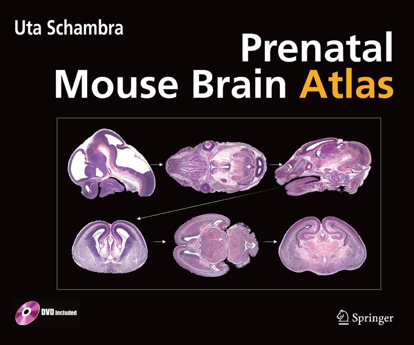
This new, greatly expanded edition presents a complete mapping of the developing mouse brain that features imaging of whole brain sections. Coverage enables users to compare structures shown in the Atlas to what they can view in a microscope.
The Prenatal Mouse Brain Atlas is the only comprehensive book available for studies of mouse brain development from early embryonic to late fetal stages. Color images of whole, hematoxylin, and eosin stained sagittal, coronal, and horizontal sections are provided at four different ages. In addition, high magnification images are included that highlight areas of developmental interest. The atlas is designed to support research of normal and abnormal brain development in developmental neuroscience, gene manipulation, molecular biology, and neurotoxicology.
Key Features:
- Color images of hematoxylin and eosin stained sections
- 26 High magnification images, highlighting areas of developmental interest
- 254 images and matching diagrams with outlined and annotated structures:
Gestational Day (GD) 12 heads:
16 GD 12 sagittal 22 GD 12 coronal 18 GD 12 horizontal
GD 14 heads:
20 GD 14 sagittal 33 GD 14 coronal 24 GD 14 horizontal
GD 16 brains:
16 GD 16 sagittal 30 GD 16 coronal 17 GD 16 horizontal
GD 18 brains: 17 GD 18 sagittal 26 GD 18 coronal 15 GD 18 horizontal
- Delineation of peripheral nerves, eyes, inner ear, ganglia and other structures in the heads of GD 12 and 14 embryos
- DVD with complete sets of images, labeled diagrams, and diagrams superimposed on images
About the author:
Dr. Uta Schambra is Associate Professor in the Department of Anatomy and Cell Biology at Quillen College of Medicine, East Tennessee State University, Johnson City, Tennessee, USA.
Inhaltsverzeichnis
Gestational Day 12 (GD 12). - Gestational Day 14 (GD 14). - Gestational Day 16 (GD 16). - Gestational Day 18 (GD 18).